

Breast health care you can trust
Freeman brings breast health services together in one place—from 3D mammograms and diagnostic imaging to Breast Lifecare, breast cancer treatment, and support with the cost of screening.
Breast cancer services
Screening options for every stage
3D mammograms
Call 417.347.7777 to schedule your mammogram today.
Low dose for diagnostic accuracy
The Senographe Pristina is the ONLY FDA approved 3-D mammography system that delivers at the same low dose as 2D full-field digital mammography, providing the lowest patient dose of all FDA approved systems.
Purpose-designed to reshape the mammography experience
The Senographe Pristina is humanizing the mammography experience by increasing comfort and reducing patient anxiety. The system’s potential to help increase the number of annual screening exams is an important step forward in women’s healthcare.
More comfortable mammography experience
Fear of pain is one of the most common reasons why women do not schedule a mammogram. A recent study conducted found that painful exams explain why 25% to 46% of women failed to return for further breast imaging. The Pristina approaches this problem in two ways: by making the paddles that compress the breast more flexible and by putting compression control in the hands of the patient.
When you compress the breast with rigid paddles in a vice, what hurts is not the compression itself but the shearing as parts of your breast move to the left and parts of your breast move to the right. GE has developed a paddle that has flexibility, resulting in much less shearing, while still giving good compression. Additionally, Pristina’s soft armrests replace the more common hand grips, helping women to relax their muscles, easing discomfort and aiding clear image acquisition.
Arkansas breast imaging and scheduling
Find breast screening and diagnostic services at our Arkansas locations. A physician order is not required for a screening mammogram. Provide a physician or qualified healthcare provider’s name for your results, and contact the imaging center about requirements for diagnostic imaging or biopsy.
Insurance coverage for 3D mammography varies. Check with your insurance provider before your appointment. Appointments are offered on a first-come, first-served basis.
Freeman Breast Imaging Center at Willow Creek
Located at Freeman Health System – Willow Creek Medical Center, the breast imaging center offers 3D mammography and upright breast biopsy using 3D technology.
For a screening mammogram, a physician order is not required. Please provide the name of the physician or qualified healthcare provider who should receive your results. If you do not have a provider, the center can provide a list to help you choose one. Reports are sent to that provider, and patients are responsible for follow-up.
Bentonville Medical Center
Freeman Health System – Bentonville Medical Center offers 3D mammography and ultrasound-guided breast biopsy. Breast tomosynthesis creates detailed images that radiologists can review one layer at a time. Ultrasound-guided biopsy uses sound waves to help guide tissue sampling from an area of concern.
Extended screening hours are available. Call the center for appointment availability, access details and any requirements for your examination.
Siloam Springs Medical Center
Freeman Health System – Siloam Springs Medical Center offers 3D mammography and upright breast biopsy using 3D technology. Stereotactic imaging helps the physician locate an area of concern for tissue sampling.
Extended screening hours are available. Call the center for appointment availability, access details and any requirements for your examination.
Fast facts
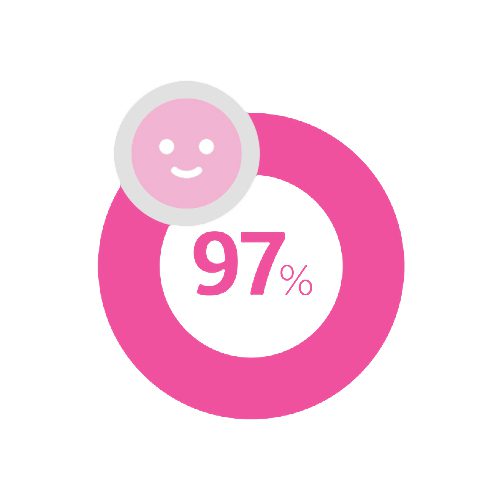
97% of patients were less anxious or not anxious at all when using Pristina.

Pristina was designed to ease patient anxiety. 50% of patients were not at all anxious during the exam.
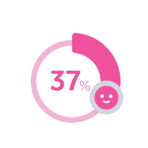
37% of patients were less anxious compared to previous exams.

First in market patient-assisted compression device
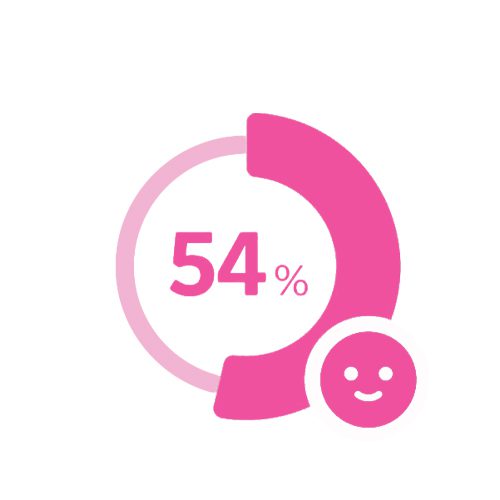
54% of patients using Dueta found it led to less anxiety.

4 out of 5 patients using Dueta said it made the exam more comfortable
FAQs

Diagnosing breast cancer
Mammography
Early detection of breast cancer greatly increases survival rates. That’s why the American College of Radiology recommends a mammogram every year for women ages 40 and older.
We can help if you do not have a physician referral for a mammogram. To schedule an appointment with Wes & Jan Houser Women’s Pavilion, call 417.347.7777.
Breast ultrasound
Breast ultrasound is a procedure used for further evaluation of an evident breast abnormality or a density-specific lump seen through mammography. It is an imaging technique using high frequency sound waves to scan the breast. Ultrasound can locate and measure abnormal changes or lesions in the breast and determine if a breast lump is solid (tumor) or filled with liquid (cyst). Ultrasound of the breast is not a replacement of mammography but rather a supplemental tool for detecting and staging breast cancer and other breast abnormalities.
Breast MRI
Magnetic resonance imaging (MRI) of the breast is a noninvasive diagnostic tool used to detect breast cancer and other abnormalities in the breast.
Breast MRI captures multiple images of the breast using a dedicated computer that generates detailed images. The study involves obtaining pictures of the breast before and after a contrast agent is administered in an effort to display not only the size and shape of a lesion, but how it enhances, which can distinguish benign and malignant lesions.
The radiologist will review the MRI and send a report to the referring physician. Breast MRI is usually performed when a physician needs more information than a mammogram, ultrasound or clinical breast exam can provide. MRI of the breast is not a replacement of mammography or ultrasound, but rather a supplemental tool for detecting and staging breast cancer and other breast abnormalities.
Learn about your biopsy
Wes & Jan Houser Women’s Pavilion uses the Mammotome® Breast Biopsy System for stereotactic breast biopsies. The system collects tissue samples through a small incision. The Mammotome system is not used for ultrasound-guided biopsies. Here’s what you can expect during a stereotactic biopsy.
Preoperative
The procedure
Postoperative
Mammotome facts
This Mammotome information is provided courtesy of Ethicon Endo-Surgery.
Breast biopsy in Arkansas
Freeman Breast Imaging Center at Willow Creek and Siloam Springs Medical Center offer upright breast biopsy using 3D technology. Patients may remain seated upright or lie on their side. The breast is compressed, as in a mammogram, while imaging helps locate the area for a small tissue sample. Your care team will explain preparation, anesthesia, recovery and how you will receive your results.
Bentonville Medical Center also offers ultrasound-guided breast biopsy, which uses sound waves to guide tissue sampling.
Arkansas biopsy contacts
For Arkansas biopsy information, contact Willow Creek at 479.757.4135, Bentonville at 479.757.4135, or Siloam Springs at 479.215.3420.
Preventing breast cancer
Steps you can take to lower your risk
Although you cannot prevent cancer, you can take steps to reduce your risk:
Early detection is the best protection
The gold standard for breast cancer screening is mammography. The American Cancer Society recommends women over age 40 have a mammogram every year. If you have a first-degree relative (mother, sister, daughter) with breast cancer, then you should start screening mammograms 10 years prior to the age she was diagnosed (but not before age 25).
Risk factors for breast cancer
Understanding your breast cancer risk
Studies have shown that your risk for breast cancer is due to a combination of factors. Not all risk factors are created equal, and having a risk factor does not mean that you will get breast cancer. The main factors that influence your risk include being a woman and getting older.
Symptoms of breast cancer
Know the signs and changes to watch for
Because breast cancer can present in many ways, and every woman’s breasts are different, patients should look for anything new or changing in the breast.
Common signs and symptoms are:
What should I do if I find a lump?
Most often, it is not cancer, but if you notice anything new or changing in your breast, reach out to your trusted care provider to get the area of concern evaluated.
Treating breast cancer
Freeman Cancer Institute provides access to cancer specialists and multiple types of treatment. Depending on your needs, your care may include surgery, chemotherapy, radiation therapy or hormone therapy. Your care team can explain the treatment options appropriate for you.
Breast Lifecare & patient navigation
Helping you find answers quickly
Whether you found a lump as result of breast self-exam, mammogram or physician’s clinical exam, the sense of urgency remains the same. You want answers and test results quickly with few delays. That’s why we developed Breast Lifecare. Our radiologists and surgeons work as a team to provide as much information as possible in as few visits as possible to minimize the stress caused by multiple appointments and waiting.
During a Breast Lifecare visit, you will have a consult with a breast surgeon. The breast surgeon will review your imaging with the breast radiologist. They will then discuss your plan of care with you. If a breast biopsy is recommended, it can be done the same day under most circumstances.
Your doctor may refer you to Breast Lifecare as a result of any of the following. If you notice any of these symptoms and do not have a current doctor, you may self-refer to the Breast Lifecare clinic by calling 417.347.7777.
You may need additional tests, which could include digital mammography, ultrasound or minimally-invasive biopsy. Most often, we can conduct these tests during your first appointment, eliminating the need for additional appointments.
Our team focuses on ensuring your comfort while delivering care in a compassionate, soothing, respectful atmosphere. With a state-of-the-art facility that features the latest technology, we take pride in providing you the best care, advice and treatment.

Our certified breast cancer patient navigator, Marcella Sowell, guides newly diagnosed breast cancer patients through the healthcare system, providing guidance from the time of diagnosis through survivorship.
With extensive experience in oncology, radiology and patient education, Sowell guides patients through the diagnostic evaluation and provides education and one-on-one support, empowering patients to make informed treatment decisions. Sowell works closely with the breast surgeons as well as medical and radiation oncologists to provide our patients with the best continuity of care, increasing patient satisfaction, safety and quality of care.
Espresso Yourself Breast Cancer Support Group meetings are also available and provide encouragement from other patients going through the same journey.
For more information about Wes & Jan Houser Women’s Pavilion, call 417.347.7777.
Arkansas breast care navigation
Dedra Walters, the Arkansas breast care nurse navigator based at Freeman Health System – Willow Creek Medical Center, supports patients and families from diagnosis through survivorship.
The navigation program provides education about diagnosis and treatment options, helps coordinate care and appointments, offers emotional support, and connects patients with community, state and national resources. Support can also include organizing health and medication information, reducing barriers to care, and helping with wig and prosthesis selection when needed. This service is provided at no cost.
Connect with Arkansas navigation
For Arkansas breast care navigation support, call 479.757.1616.
Financial assistance
Resources to make screening more accessible
Helping Friends Mammogram Fund
If you are in need of assistance for your mammogram, please mention the Helping Friends Fund while calling 417.347.7777 to schedule your mammogram.
Wes & Jan Houser Women’s Pavilion
located inside Freeman Women’s Center
1532 W. 32nd Street
Joplin, Missouri
417.347.7777
Breast Cancer Foundation of the Ozarks (BCFO)
Free screening mammograms are provided to uninsured and underinsured women who meet BCFO’s financial criteria. This program covers screening mammograms only. You can pick up an application packet at Wes & Jan Houser Women’s Pavilion front desk or contact:
Breast Cancer Foundation of the Ozarks (BCFO)
620 W. Republic Rd., Ste. 107
Springfield, MO 65807
Phone: 417.862.3838
Fax: 417.862.3830
bcfo.org
Show Me Healthy Women
Show Me Healthy Women (SMHW) offers free breast and cervical cancer screenings for Missouri women who meet age, income and insurance guidelines. The local provider can be contacted at:
Access Family Care